MBI Videos
Workshop 2: Modelling of Tissue Growth and Form
-
 Miquel Marin-Riera
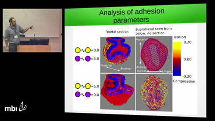
We want to understand the mechanisms that drive cell movement and tissue deformation during morphogenesis in order to predict how changes in development produce morphological variation that is relevant for evolution. We use the mammalian tooth as a model system due to the large variation it shows across the phylogenetic tree and its relatively well known development. Despite an extensive knowledge on the molecular pathways regulating the patterning and morphogenesis of the developing tooth, little is known about how individual cells move and what mechanisms are driving these movements. In order to shed light on those mechanisms, we design a mathematical model of tooth development in which cell movements are driven by compressive and tensile mechanical forces originating from tissue growth and cell-cell adhesion. The model is set to reproduce the transition between mouse molar bud (E13) and cap cap stages (E15), during which two epithelial folds protrude from the epithelial tooth germ and surround the underlying mesenchyme. When we fit the tissue specific growth rates in the model to the ones estimated from experimental data, the model correctly predicts the morphology of the tooth germ and the directionality of cell trajectories in the epithelial compartment. The model also predicts that different spatial patterns of mechanical forces arise when the adhesion strength between different cell types is varied. In order to validate the model predictions we experimentally infer the forces by means of mechanical perturbations on dissected tooth germs. We conclude that a simple model of differential growth and adhesion is able to explain the morphology, patterns of cell movement and partially the mechanical forces generated during tooth development. We argue that the addition of active cell migration might be required in order to improve the model predictions.
-
 Fengzhu Xiong
The embryonic body axis is composed of tissues that elongate at the same pace despite exhibiting strikingly different cellular organization. Whether their co-elongation is coordinated remains unclear. Here we report evidence of mechanical coupling between axial and paraxial tissues. Combining microsurgery and live-imaging in avian embryos, we found that the presomitic mesoderm (PSM) compresses the neural tube and notochord promoting their convergence and elongation. Computational simulation predicts cell motility in the PSM to generate compression that causes axial tissues to push the caudal progenitor domain, which we tested experimentally. Surprisingly, this axial push is in turn required for the progenitor addition that sustains PSM growth. Together our results show that forces produced by collective cell dynamics couple different elongating tissues into an engine-like positive feedback loop.
-
 Alexander Fletcher
Embryonic epithelia achieve complex morphogenetic movements through the coordinated action and rearrangement of individual cells. In combination with experimental approaches, computational modelling can provide insight into these processes. In this talk I describe our application of vertex models, a widely-used class of computational model for epithelia, to investigate the role of patterned cell mechanics in two settings in the Drosophila embryo: tissue size control and convergent extension. I highlight the biological insights gained through this work and conclude by presenting some recent extensions to 3D morphogenesis.
-
 Troy Shinbrot
Recent advances in the in silico modeling of cellular dynamics now permit explicit analysis of cellular structure formation. This provides the scientific community with the capability of testing hypotheses in precisely controlled environments for the first time. We describe an agent-based approach that simulates cells that reproduce, migrate and change shape. We find that both expected morphologies and previously unreported patterns spontaneously self-assemble. Most of the states found computationally have been observed in vitro, and it remains to be established what role these self-assembled states may play in in vivo morphogenesis.
-
 Hans Othmer
We will discuss how mechanics and signaling can be incorporated in cell-based models and used in a variety of contexts, including tissue growth and cell movement.
-
 Rusty Lansford
