MBI Videos
Fiber Architecture (Differential Geometry) of the Heart and its Valves
-
 Charles Peskin
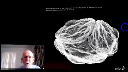
Cardiac tissue is highly anisotropic. The fibers that are responsible for this anisotropy are primarily the muscle fibers in the heart walls, and collagen fibers in the heart valve leaflets. The fiber architecture of the heart is remarkable. In the left ventricle, there are nested toroidal surfaces along which the muscle fibers run, following approximately geodesic spiral paths. In the aortic and pulmonic valves, the collagen fibers form a branching braided hammock-like structure that looks as if it might have a fractal character. The goal of the work described in this talk is to derive the fiber architecture of the heart from first principles. Our approach is to formulate partial differential equations for a system of fibers under tension supporting a pressure load, and then to see to what extent these equations predict the observed fiber architecture of the heart and its valves.
This is a National Math Biology Web Colloquium.
To watch/participate please check back here for a link on the day of the event.
